

How mitochondria affect ageing

3 mins to read

-
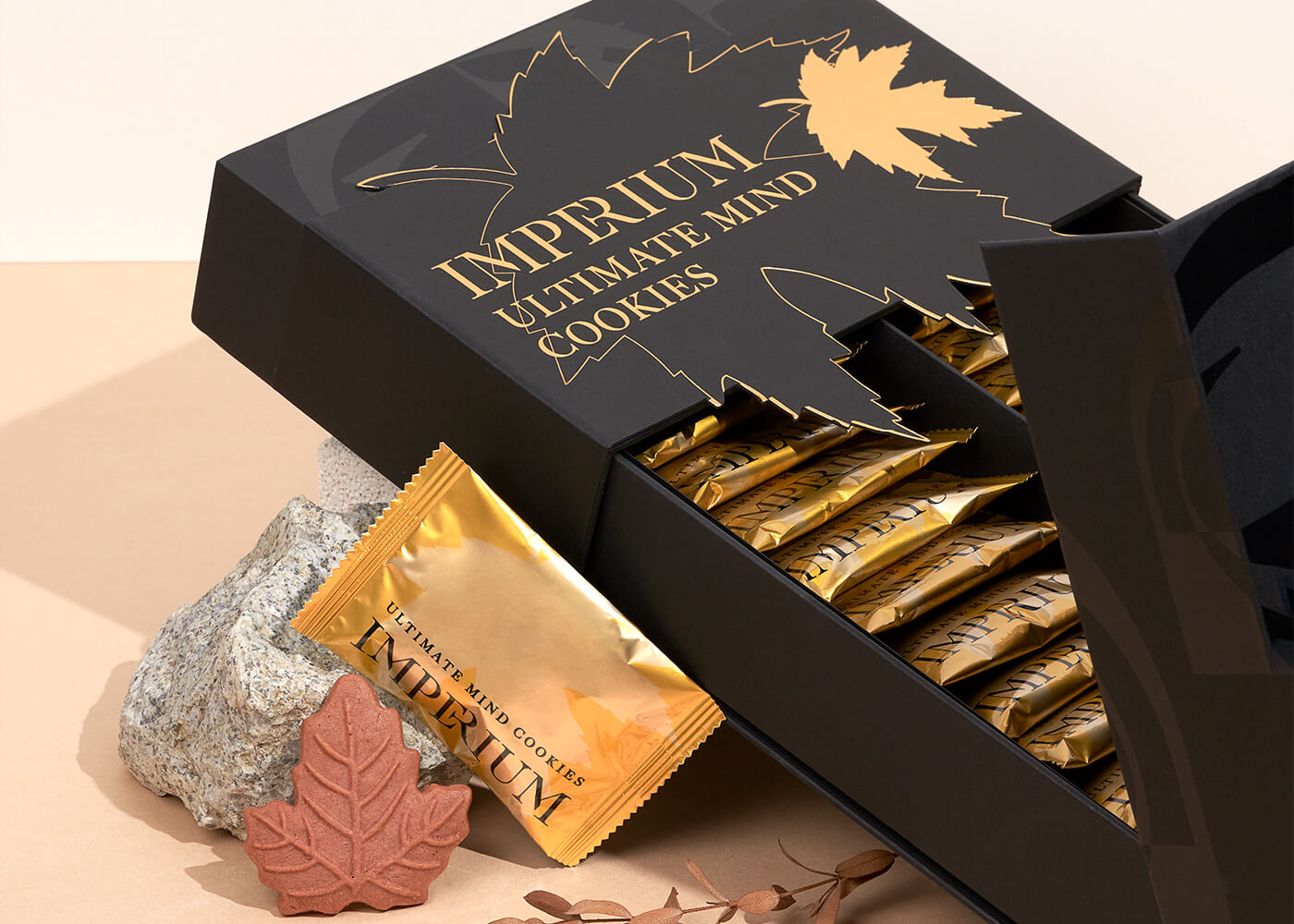
Ultimate Mind
Kick start the day with a packet of Ultimate Mind to boost memory and concentration throughout the day. The Ultimate Mind Cookie is a healthy snack that promotes memory, concentration, and cognitive function. BUY NOW How Do the Brain and Mind Function? The brain is the control centre for the body. It regulates all of […]
-

Ultimate Restore
Why is a Healthy Digestive System Important? Good digestive health is essential for good overall health. Your digestive system is responsible for breaking down the food you eat, extracting nutrients, and eliminating waste. The digestive system includes the mouth, oesophagus, stomach, small intestine, large intestine (colon), rectum, and anus. The entire length of the digestive […]
-

Ultimate Cleanse
The Ultimate Cleanse Meal Replacement is formulated with whole-food nutrition, patented and potent ingredients of more than 55 highly absorbable and functional ingredients and essential nutrients. Packed with fibre and protein, to support detoxification, cellular repair, and cellular rejuvenation. Benefits include maintaining a healthy digestive system, important for detoxification and immune health. Why is a […]